FUGUE
FUGUE is, most generally, a set of tools for EPI distortion correction. It also refers to a specific command line tool fugue.
Overview
Functional images acquired using standard EPI sequences are distorted due to magnetic field inhomogeneities.
Inhomogeneities are caused by magnetic susceptibility differences in neighbouring tissues within the head particularly for air/bone or air/tissue interfaces in the sinuses.
Inhomogeneities result in geometrical distortion and signal loss, particularly in the inferior frontal and temporal regions.
Field inhomogeneities can be measured with a fieldmap image - measured field values can be used to calculate the geometric distortion and signal loss. This information can be used to compensate for (but not completely remove) these artefacts.
Artefacts are compensated by geometrically unwarping the EPI images, and applying cost-function masking in registrations to ignore areas of signal loss.
Areas where signal loss has occurred cannot be restored with any form of post-processing, as the signal has been lost - only different acquisition techniques (which are available) can restore signal in these areas.
Distortions may be corrected for: - improving registration with non-distorted images (e.g. structurals), or - dealing with motion-dependent changes.
FUGUE is designed to deal only with the first case - improving registration. The issue of motion-dependent signal changes (due to motion-dependent changes in field inhomogeneity and distortion directions) is not dealt with in the current version.
Fieldmap Acquisition
Unfortunately, there is no standard sequence for fieldmap acquisitions and different scanners return different images. Normally these images require processing before they represent images with field values in the desired units (of radians/second) in each voxel. For SIEMENS and GEHC scanners we provide a tool: fsl_prepare_fieldmap to assist with this. Currently this does not support other scanners, but we welcome collaboration from people with other scanners to expand this functionality.
The most common fieldmap sequence acquires two images with different echo times. The change in MR phase from one image to the other is proportional to both the field inhomogeneity in that voxel and the echo time difference. The field value is therefore given by the difference in phase between these two images divided by the echo time difference. This is true for Spin Echo, Gradient Echo or EPI sequences. However, EPI-based fieldmaps suffer from the same distortions (more or less) as the functional images, while Spin Echo or Gradient Echo based fieldmap images do not. Within FSL you cannot use EPI-based fieldmaps with the standard processing, and their use in general is very problematic. We strongly recommend that Spin Echo or Gradient Echo fieldmap sequences are used to acquire the images.
MR phase is the most important quantity in a fieldmap sequence, whereas in normal imaging this phase is not of interest and is normally not saved when reconstructing the images. As a consequence, raw fieldmap scans are somewhat different from most scans, and may contain images of complex values, or separate phase and magnitude images. Furthermore, some scanners/sites may do the full reconstruction of acquired scans to yield a real-valued map of field inhomogeneities (in units of Hz, radians per second, Tesla or ppm). Alternatively no reconstruction may be done, and the raw phase and magnitude (or complex) images may be saved instead, although with modern coil arrays it can be difficult to get correct phase measurements. It is important for each different scanner/site/sequence to know what form your data is in. If they have been converted to NIFTI or ANALYZE format, then you can use the FSL tools (particularly fslinfo and FSLeyes) to determine the types of images present. To obtain fieldmaps that can be used within FSL using the FSL tools (in particular, PRELUDE and FUGUE), please refer to the section below on preparing fieldmaps for FEAT.
GUI and command line software
After you have transformed your field map into a suitable format (e.g. using fsl_prepare_fieldmap), two interfaces exist for applying FUGUE to unwarp images:
- a GUI, that is part of the FEAT preprocessing options, and
- a command-line script, epi_reg
We strongly recommend that new users make use of the pre-stats part of FEAT to do all such processing (note that it is not necessary to have fMRI data for this - even single volumes can be processed in FEAT by selecting pre-stats only). See the detailed documentation on FEAT and documentation below on making fieldmaps for FEAT.
More experienced users, or those wanting to use fieldmap-based distortion-correction in other situations, can use the epi_reg command.
Making Fieldmap Images for FEAT
This section outlines some of the most common ways to construct the required fieldmap images for B0 unwarping in FEAT. Also see the documentation on FEAT for more information on fieldmapping and its use in FEAT pre-stats processing.
Each scanner/site/sequence can give you different data. As the phase is of great importance in fieldmapping, but is normally not saved in other sequences, these images are often quite different from the standard images.
SIEMENS and GEHC data
If you have data from a SIEMENS or GEHC scanner then we strongly recommend that the tool fsl_prepare_fieldmap is used to generate the required input data for FEAT or fugue. Fieldmap data from a SIEMENS scanner takes the form of one phase difference image and two magnitude images (one for each echo time). Fieldmap data from a GEHC scanner similarly takes the form of a fieldmap in Hertz and the corresponding magnitude (See this page for more information on how to collect fieldmap data from a GEHC scanner). In the following, where a magnitude image is required, pick the "best looking" one. This image is used for registration and masking but the process is not particularly sensitive to the quality and typically either image will work fine.
There are both GUI and command-line versions of the fsl_prepare_fieldmap tool. It is recommended that new users use the GUI.
The Fsl_prepare_fieldmap GUI requires the following inputs:
- Phase image (for SIEMENS) or Fieldmap image in Hz (for GEHC)
- Magnitude image (brain extracted - see note below)
- Difference of Echo Times - this is a sequence parameter that should be in the protocol parameter PDF/printout (TE1 and TE2), if not your radiographer/ operator/technician can look this up
Brain extraction of the magnitude image is very important and must be *tight - that is, it must exclude all non-brain voxels and any voxels with only a small partial volume contribution. The reason for this is that these areas are normally very noisy in the phase image (look at them in FSLeyes - if they are not noisy then this is not so important). It is crucial that the mask (derived from this brain extracted image) contains few of these noisy voxels. This is most easily done by making the brain extraction very tight, erring on excluding brain voxels. The exclusion of brain voxels in this instance is actually fine and will have no repercussions, since the fieldmap is extrapolated beyond this mask, and that is the only purpose that the mask plays. Therefore make sure your mask is (if it can't be perfect) too small. As noted above, either magnitude image (from the different echos) can normally be used here - it is not that important.
The output of the Fsl_prepare_fieldmap GUI is a correctly calibrated fieldmap in units of rad/s (as the B0 field can be measured in Tesla, Hz or rad/s in MRI as they are all proportional).
The usage of the fsl_prepare_fieldmap command-line tool is very similar to the GUI (and all the above considerations apply):
Usage: fsl_prepare_fieldmap <scanner> <phase_image> <magnitude_image> <out_image> <deltaTE (in ms)> [--nocheck]

  Prepares a fieldmap suitable for FEAT from SIEMENS or GEHC data - saves output in rad/s format
  <scanner> must be SIEMENS or GEHC_FIELDMAPHZ
  <phase_image> should be the phase difference for SIEMENS and the fieldmap in HERTZ for GEHC_FIELDMAPHZ
  <magnitude image> should be Brain Extracted (with BET or otherwise)
  <deltaTE> is the echo time difference of the fieldmap sequence - find this out form the operator (defaults are *usually* 2.46ms on SIEMENS)
            (defaults are *usually* 2.304ms for GEHC 2D-B0MAP at 3.0T and 2.272 ms GEHC 3D B0MAP at 3.0T)
  --nocheck supresses automatic sanity checking of image size/range/dimensions

   e.g. fsl_prepare_fieldmap SIEMENS images_3_gre_field_mapping images_4_gre_field_mapping fmap_rads 2.65
   e.g. fsl_prepare_fieldmap GEHC_FIELDMAPHZ 3dB0map_fieldmaphz mag_3dB0map fmap_rads 2.272
Other data
Currently fsl_prepare_fieldmap does not work with data from other scanner manufacturers.
For users with Philips data we highly recommend looking at the following document from Anna van 't Veer, Anne Hafkemeijer, Henk van Steenbergen and Janna Marie Bas-Hoogendam (Leiden University / Leiden Institute for Brain and Cognition): https://osf.io/hks7x/ This goes through the whole process of preparing and using a fieldmap from a Philips scanner, which is hopefully standardized across scanners and sites, although at the time of writing we do not know how generally true this is.
For other scanners we welcome collaboration from people using such scanners in order to enhance the fsl_prepare_fieldmap tool.
In the meantime, when dealing with data from other scanner manufacturers, or with non-standard sequences, the first step in practice is to find out what kind of fieldmap acquisition data you can get from the scanner, operator or reconstruction software. There are three main types:
- Complex data containing images at two different echo times
- Separate phase images for the two different echo times
- A single, real fieldmap image (showing the field inhomogeneity in each voxel)
If the images from the scanner are in ANALYZE or NIFTI form then you can determine their type, use fslinfo. If not, you must convert to either ANALYZE or NIFTI format before progressing. See the FSL FAQ for information about conversion.
When fslinfo is run it will display a datatype string or number (the value 32 represents a complex image). You can convert a complex image into its phase and magnitude components by using fslcomplex or, alternatively, leave it in this format. The number of images contained in each file is shown as the dim4 value.
To further check what sort of images you have, use FSLeyes or slices to view the image and then see which of the following images it most closely resembles.
Note that for complex images, slices will display two separate images - one phase and one magnitude. If you only have a single image (dim4=1) then it is possible that it has already been reconstructed fully into a fieldmap.
Typical complex image (phase and magnitude components):
A phase image (wrapped):
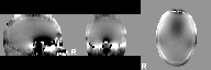
A phase image (unwrapped and masked):
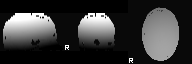
A magnitude image:
A real fieldmap image (masked):
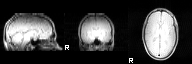
Note that the fieldmap image contains structure and shows substantial inhomogeneities mainly in the inferior regions of the brain.
It is most common to have wrapped phase and magnitude images (case 2 below). If the images appear to have unwrapped phase then the the PRELUDE stage (step 3) should be skipped. However, if the range of values within the image is within 2 pi radians (6.28) then it is likely that there are wraps present (even if they are hard to find by eye) and so, in this case, PRELUDE should still be run.
Once you have determined the type, you need to do some or all of the following steps. As a guide (and this may vary in some cases) the steps that are required are:
For complex data you need to do steps 1a, 2a, 3, 4a and 5 For a pair of phase images you need to do steps 1b, 1c, 2b, 3, 4a and 5 For a single, real fieldmap you need to do steps 1b, 1c, 4b and 5
Step 1 - Getting the magnitude image
-
(a) If you start with a complex Analyze or Nifti volume that contains the scans at two echo times then you need to do:
fslcomplex -realabs complex_acq fieldmap_mag -
(b) If you have separate phase images or a single, fieldmap image, then you need to also get a magnitude image that is (i) undistorted and (ii) registered with this phase/fieldmap image. Usually the sequence used to acquire the phase or fieldmap image also contains data that can give you this magnitude image. Check with your scanner operator, physicists and/or analysis people as to how to reconstruct this image - often it just requires extraction from the original DICOM or vendor-specific format.
-
(c) Check that the magnitude image and the phase/fieldmap images have the same resolution. You can do this by looking at the dim and pixdim entries (only the first three of each) as reported by
fslinfo.
If they are not the same then they must be resampled to be equal. In this case choose the one with the best resolution and use this as a reference image in flirt with the -applyxfm option to resample the other images. For example, if the magnitude image has a better resolution (smaller pixdims) then do the following:
    flirt -in original_phase0 -ref fieldmap_mag -applyxfm -out orig_phase0
    flirt -in original_phase1 -ref fieldmap_mag -applyxfm -out orig_phase1
Once this is done, check that the output images (e.g. orig_phase0) have the same dimensions and resolution as the reference (using fslinfo) and also check that they are aligned correctly by loading both the output and reference images into fslview and visually inspecting them.
Step 2 - Getting (wrapped) phase in radians
- (a) If you have complex volumes then do:
fslcomplex -realphase complex_acq phase0_rad 0 1
fslcomplex -realphase complex_acq phase1_rad 1 1
These phase volumes will now be in radians.
- (b) If you have seperate phase volumes that are in integer format then do:
fslmaths orig_phase0 -mul 3.14159 -div 2048 phase0_rad -odt float
fslmaths orig_phase1 -mul 3.14159 -div 2048 phase1_rad -odt float
Note that the value of 2048 needs to be adjusted for each different site/scanner/sequence in order to be correct. The final range of the phase0_rad image should be approximately 0 to 6.28. If this is not the case then this scaling is wrong. If you have separate phase volumes are not in integer format, you must still check that the units are in radians, and if not scale them appropriately using fslmaths.
Step 3 - Unwrapping the phase images
Use prelude to do the required phase unwrapping
prelude -a fieldmap_mag -p phase0_rad -o phase0_unwrapped_rad
prelude -a fieldmap_mag -p phase1_rad -o phase1_unwrapped_rad
Step 4 - Getting the fieldmap in rad/s
- (a) For separate phase images do:
fslmaths phase1_unwrapped_rad -sub phase0_unwrapped_rad -mul 1000 -div <TE> fieldmap_rads -odt float
where <TE> must be replaced with the appropriate difference in echo times (in units of milliseconds).
- (b) If you have a single, real fieldmap then you must determine the units of this fieldmap (ask an operator/physicist) and rescale to radians per second if it is not already in these units. Common other units are (i) Hz (scale these by 6.28 to get rad/s) and (ii) Telsa (scale these by 2.68e8 to get rad/s).
Step 5 - Regularising the fieldmap
Fieldmaps can often be noisy or be contaminated around the edges of the brain. To correct for this you can regularise the fieldmap using fugue. Note that the "best" regularisation will depend on many factors in the acquisition and must be determined separately for each site/scanner/sequence. Look at the fieldmap (e.g. using FSLeyes) to decide what is the best regularisation to use - which could also be to do no regularisation.
Some regularisation options are - Gaussian smoothing, despiking and median filtering. Examples of these (in order) are:
fugue --loadfmap=fieldmap_rads -s 1 --savefmap=fieldmap_rads
fugue --loadfmap=fieldmap_rads --despike --savefmap=fieldmap_rads
fugue --loadfmap=fieldmap_rads -m --savefmap=fieldmap_rads
Any combination of these regularisations can be performed. See the command-line documentation on fugue below for more information aspects of regularisation.
Command-line tools (advanced)
For all programs the options follow the normal convention that "single minus" options are separated by a space from their arguments (if any) whilst "double minus" options are separated by an equals sign and no space. For example,
prelude -c data --unwrap=result
or
prelude --complex=data -u result
fugue (EPI unwarping)
fugue (FMRIB's Utility for Geometrically Unwarping EPIs) performs unwarping of an EPI image based on fieldmap data.
The input required consists of the EPI image, the fieldmap (as an unwrapped phase map or a scaled fieldmap in rad/s) and appropriate image sequence parameters for the EPI and fieldmap acquisitions: the dwell time for EPI (also known as the echo spacing); and the echo time difference (called asym time herein).
The main forms of usage are:
-
With the fieldmap specified by a 4D file containing two unwrapped phase images - from different echo times - plus the dwell time and echo time difference (asym time):
fugue -i epi -p unwrappedphase --dwell=dwelltime --asym=asymtime -s 0.5 -u result -
Using a previously calculated fieldmap:
fugue -i epi --dwell=dwelltime --loadfmap=fieldmap -u result
Note the option -s 0.5 is an example of how to specify the regularisation to apply to the fieldmap (2D Gaussian smoothing of sigma=0.5 in this case which is a reasonable default). There are many different forms of regularisation available which can be applied separately or together. These are:
-s sigma: 2D Gaussian smoothing--smooth3=sigma: 3D Gaussian smoothing-m: 2D median filtering--poly=n: 3D Polynomial fitting of degree n--fourier=n: 3D Sinusoidal fitting of degree n
Some other uses are:
fugue -i undistortedimage -p unwrappedphase --dwell=dwelltime --asym=asymtime --nokspace -s 0.5 -w warpedimage
applies the fieldmap as a forward warp, turning an undistorted image into a distorted one - useful for creating a registration target for the EPI from the undistorted absolute fieldmap image.
Additional options that are useful are as follows (run fugue without any arguments for details on all options):
-
--mask=maskname: uses a user-defined mask (calledmaskname) instead of deriving it from the phasemaps or fieldmaps -
--unwarpdir=dir: specifies the direction of the unwarping/warping - i.e. phase-encode direction - withdirbeing one ofx,y,z,x-,y-,z-(default isy).Note: the conventions for x, y and z are voxel axes (in the input image), but with the x-axis being swapped if the qform or sform determinant is positive (to match the internal FSL voxel coordinate axes), though due to other possible changes in the pipeline for reconstructing/analysing both the EPI and fieldmap images, it is usually necessary to determine the sign of the direction by trial-and-error once for each scanning/reconstruction/analysis protocol (each dataset using the same protocol will need the same unwarpdir).
-
--phaseconj: uses the phase conjugate correction method, rather than pixel shifts -
--nokspace: for forward warping (only) - uses an image-space method for forward warping -
--icorr: applies an intensity correction term when using the pixel shift method - often poorly conditioned for standard fieldmap acquisitions
prelude (phase unwrapping)
prelude (Phase Region Expanding Labeller for Unwrapping Discrete Estimates) performs 3D phase unwrapping of images. As the name implies, it should be run before fugue in order to get a correct fieldmap. This is useful for fieldmap acquisitions, susceptibility weighted imaging (SWI), or other applications involving phase in MR (or non-MR) images. The input can either be a single complex image (NIfTI or Analyze), or a pair of real images giving the phase and absolute values separately. Also see the fslcomplex utility for more ways to manipulate complex image formats. If the images are 4D images, then each 3D volume is unwrapped separately, and the result saved as a 4D image of unwrapped phase images. The output in either case is a real, unwrapped phase image (in radians).
For higher resolution images (e.g. for Susceptibility Weighted Imaging) a mask should be used in order to reduce run-times since any areas that include noisy voxels will significantly increase the required run-time. The best way to generate a mask for a whole brain image is to run BET to perform brain extraction on the absolute image.
The three main forms of usage are:
-
Using a single complex input file:
prelude -c data -u result -
Using separate phase and absolute input files:
prelude -a data_abs -p data_phase -u result -
Using separate phase and absolute input files with a mask:
prelude -a data_abs -p data_phase -u result -m mask
Additional options that are useful are (run prelude without any arguments for details on all options):
-
-m mask: uses the user-defined mask (e.g. a BET mask - especially for high resolution images) -
-n num: specifies the number of phase partitions the algorithm uses for initial labelling - a larger value is likely to be more robust but slower -
-s: unwrap in 2D, then stick slices together with appropriate offsets - this is less robust but fast - mainly used for very high-resolution images where optimising speed is an issue -
--labelslices: does labelling in 2D, but unwrapping in 3D - the default for high-res images
FAQ
What type of field maps do I need for (PRELUDE and) FUGUE?
The fieldmaps required by FUGUE must be in undistorted coordinates. That is, the field map should not have any geometric distortion in it (or negligible amounts) as can be obtained from a spin-echo sequence, or gradient-echo sequences if the echo time is sufficiently small. The distortion present in a sequence can readily be seen in the absolute (or magnitude) image and normally manifests itself as a depression or extension of the inferior frontal lobes (when the scans are acquired axially - it is different for other acquisition planes). It is recommended that a symmetric-asymmetric spin-echo sequence is used for the field mapping. This sequence acquires one conventional spin-echo image and then another with the same settings except with the 180-degree RF pulse offset by a small delay, known as the asym-time. The difference in the phase of the two images is then proportional to the field offset.
Note that the input to PRELUDE, for phase unwrapping, must be a complex image either presented as a single 3D or 4D complex Analyze image or as a set of two 3D or 4D Analyze images - one for the phase and one for the absolute (or magnitude) part. The input to FUGUE must be a 4D Analyze image containing two unwrapped phase maps. To convert between 3D and 4D Analyze files the utilities fslmerge and fslsplit can be used. To convert between pairs of real images and complex images, the utility fslcomplex can be used.
How do I get a fieldmap sequence for my scanner?
We cannot provide field-map sequences for different scanners. The recommended sequence is a symmetric-asymetric spin-echo sequence as described above. If such a sequence is not available at your site, try contacting the FSL email list to see if others have a sequence for your scanner that could be used.