In vivo MRI
The animal was cared for, and data were acquired by researchers at the University of Oxford, UK. All procedures were performed under licenses from the United Kingdom (UK) Home Office in accordance with the UK Animals (Scientific Procedures) Act 1986 and with European Union guidelines (EU Directive 2010/63/EU).
In vivo data were acquired across multiple scanning sessions. Here we provide resting-state fMRI, structural and diffusion MRI data from two scanning sessions (named “prime-de”/”last-scan”), ~7 years apart. Some of the data (rs-fmri & structural) from the first session are already available via the primate data exchange (PRIME-DE) where complementary data from another 19 subjects is available (Noonan 2014). Data from the second session constitutes the last in vivo data acquired, approximately 1 year prior to ex vivo data acquisition (“last-scan”).
Task-fMRI data were also acquired. Z-stat maps linking behaviour to fMRI are available via Grohn 2020 (doi:10.1371/journal.pbio.3000899) and Sallet 2020 (doi:10.1371/journal.pbio.3000605). Full references are provided at the bottom of this page.
Data were acquired on a 3T whole-body scanner with 40 G/cm gradients, a four-channel phased array receive coil and a local transmit coil. More information about the acquisition procedures can be found at https://fcon_1000.projects.nitrc.org/indi/PRIME/oxford.html.
The data were preprocessed using FSL tools built into bespoke macaque pipelines (MrCat: https://github.com/neuroecology/MrCat).
Structural MRI
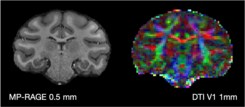
T1-weighted data at 0.5 mm isotropic resolution were acquired using an MP-RAGE sequence.
MP-RAGE acquisition parameters: TE/TR = 4.01 ms/2.5 s; 128 slices; 0.5 mm isotropic resolution; 5/2 images (repeats) were acquired for the first and second dataset (prime-de/last-scan) respectively.
Diffusion MRI
Diffusion MRI were acquired at 1 mm isotropic resolution using echo planar imaging.
EPI acquisition parameters: TE/TR = 100 ms/8.2 s; b-value = 1 ms/μm2; 1 mm isotropic resolution; 1100/361 diffusion weighted (81/61 unique gradient directions) and 144/38 non-diffusion weighted volumes were acquired with both +/- phase encoding directions for the first and second dataset (prime-de/last-scan) respectively.
Resting-state fMRI
Resting-state functional MRI data were acquired using EPI at 2 mm isotropic resolution.
EPI acquisition parameters: TE/TR = 19 ms/2 s; 1600/800 volumes for the first and second dataset (prime-de/last-scan) respectively. This corresponds to 52 min 26 s or 26 min 13 s of data.
Below are examples images of the bias-field corrected structural MRI and the primary eigenvector of the diffusion tensor (DTI V1) from the “prime-de” dataset.
Key references:
Noonan, M.A.P., Sallet, J., Mars, R.B., Neubert, F.X., O’Reilly, J.X., Andersson, J.L., Mitchell, A.S., Bell, A.H., Miller, K.L., Rushworth, M.F.. A Neural Circuit Covarying with Social Hierarchy in Macaques. PLOS Biology 2014;12(9):e1001940. doi:10.1371/journal.pbio.1001940.
Grohn, J., Schuffelgen, U., Neubert, F.X., Bongioanni, A., Verhagen, L., Sallet, J., Kolling, N., Rushworth, M.F.. Multiple systems in macaques for tracking prediction errors and other types of surprise. PLOS Biology 2020;18(10):e3000899. doi:10.1371/journal.pbio.3000899.
Sallet, J., Noonan, M.A.P., Thomas, A., O’Reilly, J.X., Anderson, J., Papageorgiou, G.K., Neubert, F.X., Ahmed, B., Smith, J., Bell, A.H., Buckley, M.J., Roumazeilles, L., Cuell, S., Walton, M.E., Krug, K., Mars, R.B., Rushworth, M.F.. Behavioral flexibility is associated with changes in structure and function distributed across a frontal cortical network in macaques. PLoS Biology 2020;18(5):e3000605. doi:10.1371/journal.pbio.3000605.